
WORLD
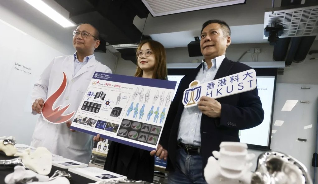
Hong Kong researchers have developed artificial intelligence (AI) technology that generates 3D bone and organ models for implants from X-ray images, which they claim reduces patients’ radiation exposure by up to 99 per cent compared to computed tomography (CT) scans.
The team at Hong Kong University of Science and Technology (HKUST) said they hoped that the technology would be adopted in the city’s public healthcare system as it could lead to substantial cost savings and shorter wait times for generating 3D medical models.
Currently, medical professionals rely on CT scans to produce 3D models of bones and organs for patients. However, this conventional method emits high levels of radiation, posing potential risks, especially to vulnerable groups such as children, pregnant women, and elderly patients who require frequent monitoring.
“CT imaging is widely used in medicine, but it has drawbacks, such as high radiation levels, lack of portability, and long waiting times for patients to undergo CT scans in Hong Kong,” said Professor Li Xiaomeng, assistant professor from the Department of Electronic and Computer Engineering at HKUST.
“X-rays are cost-effective alternatives, but their limitation is the inability to achieve high-definition 3D images. Our AI technology compensates for this weakness.”
Li, who is also the associate director of the university’s Centre for Medical Imaging and Analysis, said 500 patient cases were used to train the AI model and 120 sets of data to compare its accuracy to that of CT scans.
She added that with the new AI-driven technology, doctors could upload two to four X-ray images to a platform to generate a 3D bone model in under 30 seconds, compared to 400 to 500 X-ray snapshots typically required for a CT scan, thus cutting radiation exposure by 95 to 99 per cent.
She also said the innovation had an average surface distance error of less than one millimetre, making it the first of its kind to achieve such precision.
Other than bone models, it was expected that the technology could also be applied to teeth and lung imaging.
Li’s team collaborated with Koln 3D, Hong Kong’s first orthopaedic and metal component printing technology company, to apply the technology in pre-surgical planning, for personalised and in real-time surgical navigation.
Edmond Yau Wing-fung, founder and CEO of Koln 3D, said without the technology, doctors and hospitals would have to pay an expensive fee for software that was the only internationally approved solution in the market that could turn CT scan images into 3D models.
But he said that the software “was not user-friendly” and required five to six hours to complete the process.
He added that a local public hospital would further test the technology’s accuracy, with the expectation that it could be used for printing 3D implants for surgeries within a year.
Currently, processing CT scans for designing and printing 3D implants can take hours.
Dr Tse Lung-fung, a specialist in orthopaedics and traumatology, said the technology had significant potential for applications such as correcting limb deformities requiring surgical intervention and treating trauma conditions like bone fractures that need surgical fixation.
He added that patients would only need to pay HK$300 (US$38) to HK$400 for an X-ray, while a CT scan would cost around HK$3,000 to $4,000.
Furthermore, patients would be spared the lengthy waiting lists for CT scans at public hospitals, which can typically take three weeks.
“CT scans are sometimes required during surgeries, but medical professionals can be exposed to a high level of radiation, which can be worrying,” he said.
“The safety of patients and medical professionals is crucial to us.”
He added that doctors from France and Singapore had also expressed interest in the technology. – SCMP
Leave a Reply